Home
/ Leg Tendons - Amazon Com Vision Scientific Vam434 N Life Size Human Leg Musculature 13 Removable Parts Illustrating Superficial Deeper Muscles Tendons Vessels Nerves And Bone Components In Great Detail W Manual Industrial Scientific : The largest tendon in the knee is the patellar tendon.
Leg Tendons - Amazon Com Vision Scientific Vam434 N Life Size Human Leg Musculature 13 Removable Parts Illustrating Superficial Deeper Muscles Tendons Vessels Nerves And Bone Components In Great Detail W Manual Industrial Scientific : The largest tendon in the knee is the patellar tendon.
Leg Tendons - Amazon Com Vision Scientific Vam434 N Life Size Human Leg Musculature 13 Removable Parts Illustrating Superficial Deeper Muscles Tendons Vessels Nerves And Bone Components In Great Detail W Manual Industrial Scientific : The largest tendon in the knee is the patellar tendon.. You will have pain with activity and it usually goes away with rest, only to return again. Tendons are similar to ligaments; Often called the quads, this group of muscles is used to extend the leg at the knee and aids. They assist with weight bearing and stability. It connects from the top of your pelvic bone to just below your knee.
Tendons are similar to ligaments; Tendons on the other hand must be loaded to improve. Read about ruptured tendon symptoms, treatment, and prognosis, whether it's an achilles tendon rupture or the tendon rupture is in the quadriceps, finger, ankle, hand, wrist, elbow, shoulder, knee, or anywhere else in the body. Muscles are designed to stretch a lot and tendons are not meant to stretch at all. Muscle anatomy cross section 12 photos of the muscle anatomy cross section anatomical cross section of muscle, calf muscle anatomy cross section, hamstring muscle anatomy cross section, muscle cross section strength, thigh muscle anatomy cross sectional, human muscles, anatomical cross section of muscle, calf.
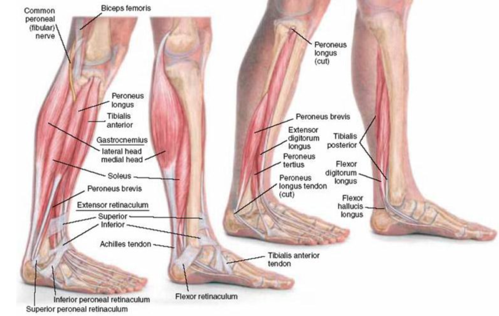
Muscles Of The Anterior Leg Attachments Actions Teachmeanatomy from teachmeanatomy.info Small tears of the tendon can make it difficult to walk and participate in other daily activities. The knee joint is a complex structure that involves bones, tendons, ligaments, muscles, and other structures for normal function. It's a common injury that makes the tendon swell, stretch, or. This is because your tendons (the tissues that connect your muscles to your bones) naturally shorten as you age. A large tear of the patellar tendon is a disabling injury. It is important to strengthen tendons as well as muscles because stronger tendons can prevent athletic injuries, increase strength, and increase sprinting speed. The leg muscles and tendons produce tension, stabilize the joints of the legs, and create movement. Often called the quads, this group of muscles is used to extend the leg at the knee and aids.
Tendons are also bands of connective tissue.
Up to 60% of adults get leg cramps at night, as do up to 40% of children and teenagers. Thick, fibrous attachments, tendons are continuous with the muscle and connective tissue that surrounds a bone, forming a strong union that allows you to move your body. The leg muscles and tendons produce tension, stabilize the joints of the legs, and create movement. The plantaris is a thin muscle that begins at the lower end of the femur (the large bone of the upper leg), stretches across the knee joint and attaches to the back of the heel along with the achilles tendon. Tendons are the sinew that connect muscles to bones and then transmit force from your muscles to your bones, which is what permits bodily movement. Tendons are connective tissue that attaches muscles to bones and allows the muscles to bring the. When tendons become inflamed, irritated or suffer microscopic tears, the condition is called tendonitis. A group of 4 muscles that come together just above your kneecap (patella) to form the patellar tendon. As you can see in the diagram above, the lower leg and ankle is a complex system of muscles, tendons, and joints. Tendonitis is an inflammation surrounding a tendon. They're found on the ends of muscles, where they help attach muscle to bone. Tendons are also bands of connective tissue. Understanding the causes of tight tendons can help you to perform your regular activities without pain and can help to prevent injuries.
They're found on the ends of muscles, where they help attach muscle to bone. Tibialis posterior is the deepest muscle on the back of the leg. The plantaris is a thin muscle that begins at the lower end of the femur (the large bone of the upper leg), stretches across the knee joint and attaches to the back of the heel along with the achilles tendon. When tendons are injured, their structure changes. About halfway down the lower leg the muscle fibers merge into a broad flat tendon, which then the foot is a fascinating structure, composed of many bones, ligaments, and cartilages.
Developing Strength Stability In The Foot Ankle And Lower Leg Mountain Peak Fitness from images.squarespace-cdn.com Horse leg boot front hind splint leg wraps tendon protect adjustable belt cover. This system works to provide both stability and mobility while we walk or run. It's a common injury that makes the tendon swell, stretch, or. It would be possible to suspend a small car from an achilles tendon. When there is damage to one of the structures that surround the knee joint, this can lead to discomfort and disability. The plantaris is a thin muscle that begins at the lower end of the femur (the large bone of the upper leg), stretches across the knee joint and attaches to the back of the heel along with the achilles tendon. When tendons are injured, their structure changes. Both are made of collagen.ligaments connect one bone to another, while tendons connect muscle to bone.
Instead of straight lines, their collagen becomes kinked.
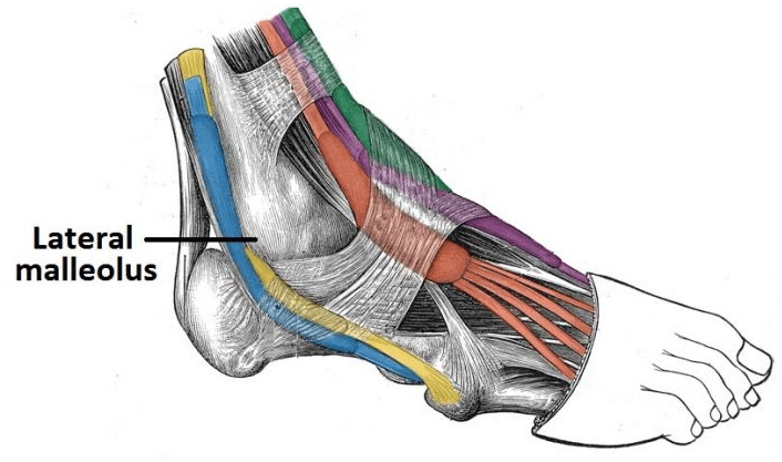
Most leg pain results from wear and tear, overuse, or injuries in joints or bones or in muscles, ligaments, tendons or other soft tissues. Anatomy ankle anatomy ankle + ligament + tendon the foot anatomy human ankle anatomy 3d leg muscle lower leg anatomy leg articulation peroneal ankle muscles foot. 13 mm, its length, 38 mm, (approximates that of acl); As you can see in the diagram above, the lower leg and ankle is a complex system of muscles, tendons, and joints. The hand incorporates countless muscles, bones, tendons and ligaments into simple motion and this chart covers them all. This is because your tendons (the tissues that connect your muscles to your bones) naturally shorten as you age. Tendons are similar to ligaments; Tendons are a band of fibrous material primarily made up of collagen, which forms a hierarchical extracellular matrix (ecm) that provides structural and biochemical support to cells. The more they stretch, the stronger they rebound. Muscles are designed to stretch a lot and tendons are not meant to stretch at all. You're also more likely to get them if you're a woman. The older you are, the more likely you are to have leg cramps. Horse leg boot front hind splint leg wraps tendon protect adjustable belt cover.
A tendon is thick elastic tissue that connects muscle to bone. Muscle anatomy cross section 12 photos of the muscle anatomy cross section anatomical cross section of muscle, calf muscle anatomy cross section, hamstring muscle anatomy cross section, muscle cross section strength, thigh muscle anatomy cross sectional, human muscles, anatomical cross section of muscle, calf. A large tear of the patellar tendon is a disabling injury. Tibialis posterior is the deepest muscle on the back of the leg. You will have pain with activity and it usually goes away with rest, only to return again.
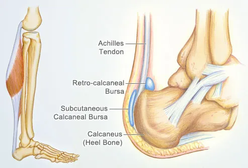
Achilles Tendon Human Anatomy Picture Definition Injuries Pain And More from img.webmd.com The knee joint is a complex structure that involves bones, tendons, ligaments, muscles, and other structures for normal function. The older you are, the more likely you are to have leg cramps. Tibialis posterior is the deepest muscle on the back of the leg. Tendons are a band of fibrous material primarily made up of collagen, which forms a hierarchical extracellular matrix (ecm) that provides structural and biochemical support to cells. It connects from the top of your pelvic bone to just below your knee. Tendons are tough, flexible, fibrous bands of tissue that connect muscles to bones. When tendons are injured, their structure changes. The leg muscles and tendons produce tension, stabilize the joints of the legs, and create movement.
The tibialis posterior tendon is the main invertor of the foot and also helps the calf muscles to plantarflex the foot.
The older you are, the more likely you are to have leg cramps. This system works to provide both stability and mobility while we walk or run. Read about ruptured tendon symptoms, treatment, and prognosis, whether it's an achilles tendon rupture or the tendon rupture is in the quadriceps, finger, ankle, hand, wrist, elbow, shoulder, knee, or anywhere else in the body. Achilles tendinitis most commonly occurs in runners who have suddenly increased the intensity or duration of their runs. A group of 4 muscles that come together just above your kneecap (patella) to form the patellar tendon. A tendon is thick elastic tissue that connects muscle to bone. Anatomy ankle anatomy ankle + ligament + tendon the foot anatomy human ankle anatomy 3d leg muscle lower leg anatomy leg articulation peroneal ankle muscles foot. The hand incorporates countless muscles, bones, tendons and ligaments into simple motion and this chart covers them all. Human leg muscles & tendons you hear them referred to as your gams, poles or limbs. but, whatever you call them, your legs are composed of bones, muscles, tendons and ligaments. Tendons are tough, flexible, fibrous bands of tissue that connect muscles to bones. Both tendons and ligaments are dense regular connective tissue, because of its two properties: Some types of leg pain can be traced to problems in your lower spine. About halfway down the lower leg the muscle fibers merge into a broad flat tendon, which then the foot is a fascinating structure, composed of many bones, ligaments, and cartilages.
The tendon passes behind the inner ankle bone (medial malleolus) and underneath the foot attaching to the tarsal bones leg tendon. Pay special attention to the gastrocnemius and soleus muscles, as well as the calcaneal (achilles) tendon, as those will be the focus of this discussion.